Application of modern autoradiography to nuclear forensic analysis
- Lawrence Livermore National Lab. (LLNL), Livermore, CA (United States)
- Lawrence Livermore National Lab. (LLNL), Livermore, CA (United States); Univ. of Nevada, Las Vegas, NV (United States)
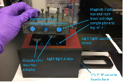
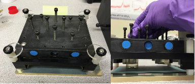
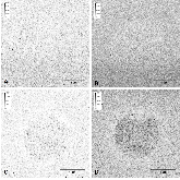
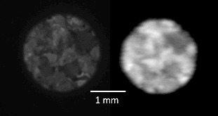
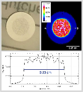
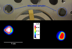
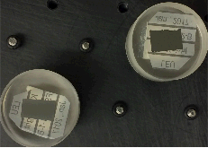
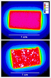
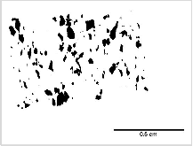
Modern autoradiography techniques based on phosphorimaging technology using image plates (IPs) and digital scanning can identify heterogeneities in activity distributions and reveal material properties, serving to inform subsequent analyses. Here, we have adopted these advantages for applications in nuclear forensics, the technical analysis of radioactive or nuclear materials found outside of legal control to provide data related to provenance, production history, and trafficking route for the materials. IP autoradiography is a relatively simple, non-destructive method for sample characterization that records an image reflecting the relative intensity of alpha and beta emissions from a two-dimensional surface. Such data are complementary to information gathered from radiochemical characterization via bulk counting techniques, and can guide the application of other spatially resolved techniques such as scanning electron microscopy (SEM) and secondary ion mass spectrometry (SIMS). IP autoradiography can image large 2-dimenstional areas (up to 20 × 40 cm), with relatively low detection limits for actinides and other radioactive nuclides, and sensitivity to a wide dynamic range (105) of activity density in a single image. Distributions of radioactivity in nuclear materials can be generated with a spatial resolution of approximately 50 μm using IP autoradiography and digital scanning. While the finest grain silver halide films still provide the best possible resolution (down to ~10 μm), IP autoradiography has distinct practical advantages such as shorter exposure times, no chemical post-processing, reusability, rapid plate scanning, and automated image digitization. Sample preparation requirements are minimal, and the analytical method does not consume or alter the sample. These advantages make IP autoradiography ideal for routine screening of nuclear materials, and for the identification of areas of interest for subsequent micro-characterization methods. Here in this article we present a summary of our setup, as modified for nuclear forensic sample analysis and related research, and provide examples of data from select samples from the nuclear fuel cycle and historical nuclear test debris.
- Research Organization:
- Lawrence Livermore National Laboratory (LLNL), Livermore, CA (United States)
- Sponsoring Organization:
- USDOE; USDHS
- Grant/Contract Number:
- AC52-07NA27344
- OSTI ID:
- 1438774
- Report Number(s):
- LLNL-JRNL-735865; TRN: US1900521
- Journal Information:
- Forensic Science International, Vol. 286, Issue C; ISSN 0379-0738
- Publisher:
- ElsevierCopyright Statement
- Country of Publication:
- United States
- Language:
- English
Web of Science
Isotopic and Compositional Variations in Single Nuclear Fuel Pellet Particles Analyzed by Nanoscale Secondary Ion Mass Spectrometry
|
journal | December 2019 |
Similar Records
Quantitative single-particle digital autoradiography with α-particle emitters for targeted radionuclide therapy using the iQID camera
Quantitative Digital Autoradiography for Environmental Swipe Sample Prioritization: System design, Characterization, and Initial Measurements