
Polarization induced contrast X-ray fluorescence at submicrometer resolution reveals nanometer apatite crystal orientations across entire tooth sections
Abstract
For biomedical research, successful imaging of calcified microstructures often relies on absorption differences between features, or on employing dies with selective affinity to areas of interest. When texture is concerned, e.g. for crystal orientation studies, polarization induced contrast is of particular interest. This requires sufficient interaction of the incoming radiation with the volume of interest in the sample to produce orientation-based contrast. Here we demonstrate polarization induced contrast at the calcium K-edge using submicron sized monochromatic synchrotron X-ray beams. We exploit the orientation dependent subtle absorption differences of hydroxyl-apatite crystals in teeth, with respect to the polarization field of the beam. Interaction occurs with the fully mineralized samples, such that differences in density do not contribute to the contrast. Our results show how polarization induced contrast X-ray fluorescence mapping at specific energies of the calcium K-edge reveals the micrometer and submicrometer crystal arrangements in human tooth tissues. This facilitates combining both high spatial resolution and large fields of view, achieved in relatively short acquisition times in reflection geometry. In enamel we observe the varying crystal orientations of the micron sized prisms exposed on our prepared surface. We easily reproduce crystal orientation maps, typically observed in polished thin sections. We evenmore »
- Publication Date:
- Research Org.:
- Lawrence Livermore National Lab. (LLNL), Livermore, CA (United States)
- Sponsoring Org.:
- USDOE National Nuclear Security Administration (NNSA)
- OSTI Identifier:
- 1484326
- Alternate Identifier(s):
- OSTI ID: 1497983
- Report Number(s):
- LLNL-JRNL-756138
Journal ID: ISSN 2156-7085
- Grant/Contract Number:
- AC52-07NA27344
- Resource Type:
- Published Article
- Journal Name:
- Biomedical Optics Express
- Additional Journal Information:
- Journal Name: Biomedical Optics Express Journal Volume: 10 Journal Issue: 1; Journal ID: ISSN 2156-7085
- Publisher:
- Optical Society of America
- Country of Publication:
- United States
- Language:
- English
- Subject:
- 36 MATERIALS SCIENCE; 47 OTHER INSTRUMENTATION; 59 BASIC BIOLOGICAL SCIENCES
Citation Formats
                    Hesse, Bernhard, Stier, Deborah, Cotte, Marine, Forien, Jean-Baptiste, and Zaslansky, Paul. Polarization induced contrast X-ray fluorescence at submicrometer resolution reveals nanometer apatite crystal orientations across entire tooth sections.  United States: N. p., 2018.
Web.  doi:10.1364/BOE.10.000018.
Hesse, Bernhard, Stier, Deborah, Cotte, Marine, Forien, Jean-Baptiste, & Zaslansky, Paul. Polarization induced contrast X-ray fluorescence at submicrometer resolution reveals nanometer apatite crystal orientations across entire tooth sections. United States. https://doi.org/10.1364/BOE.10.000018
                    Hesse, Bernhard, Stier, Deborah, Cotte, Marine, Forien, Jean-Baptiste, and Zaslansky, Paul. Mon .
"Polarization induced contrast X-ray fluorescence at submicrometer resolution reveals nanometer apatite crystal orientations across entire tooth sections".  United States.  https://doi.org/10.1364/BOE.10.000018.
@article{osti_1484326,

  title        = {Polarization induced contrast X-ray fluorescence at submicrometer resolution reveals nanometer apatite crystal orientations across entire tooth sections},

  author       = {Hesse, Bernhard and Stier, Deborah and Cotte, Marine and Forien, Jean-Baptiste and Zaslansky, Paul},

  abstractNote = {For biomedical research, successful imaging of calcified microstructures often relies on absorption differences between features, or on employing dies with selective affinity to areas of interest. When texture is concerned, e.g. for crystal orientation studies, polarization induced contrast is of particular interest. This requires sufficient interaction of the incoming radiation with the volume of interest in the sample to produce orientation-based contrast. Here we demonstrate polarization induced contrast at the calcium K-edge using submicron sized monochromatic synchrotron X-ray beams. We exploit the orientation dependent subtle absorption differences of hydroxyl-apatite crystals in teeth, with respect to the polarization field of the beam. Interaction occurs with the fully mineralized samples, such that differences in density do not contribute to the contrast. Our results show how polarization induced contrast X-ray fluorescence mapping at specific energies of the calcium K-edge reveals the micrometer and submicrometer crystal arrangements in human tooth tissues. This facilitates combining both high spatial resolution and large fields of view, achieved in relatively short acquisition times in reflection geometry. In enamel we observe the varying crystal orientations of the micron sized prisms exposed on our prepared surface. We easily reproduce crystal orientation maps, typically observed in polished thin sections. We even reveal maps of submicrometer mineralization fronts in spherulites in intertubular dentine. This Ca K-edge polarization sensitive method (XRF-PIC) does not require thin samples for transmission nor extensive sample preparation. It can be used on both fresh, moist samples as well as fossilized samples where the information of interests lies in the crystal orientations and where the crystalline domains extend several micrometers beneath the exposed surface.},

  doi          = {10.1364/BOE.10.000018},

  journal      = {Biomedical Optics Express},

  number       = 1,

  volume       = 10,

  place        = {United States},

  year         = {Mon Dec 03 00:00:00 EST 2018},

  month        = {Mon Dec 03 00:00:00 EST 2018}

}
https://doi.org/10.1364/BOE.10.000018
 Search WorldCat to find libraries that may hold this journal
Web of Science
Figures / Tables:
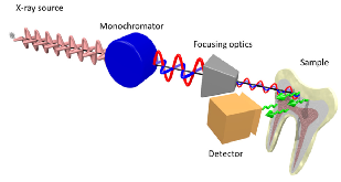 Figure 1: Schematic illustration of the main optical components of the setup installed at beamline ID21 for XRF-PIC mapping. Two undulators are used to generate the X-rays that are polarized in the horizontal plane, subsequently monochromatized by a fixed exit double-crystal Si(111) Kohzu-monochromator, then focused using KB optics. The polarizationmore »
Works referenced in this record:
Dentin structure composition and mineralization
journal, January 2011
- Goldberg, Michel
- Frontiers in Bioscience, Vol. E3, Issue 2
Polarization Dependence of Aragonite Calcium L-Edge XANES Spectrum Indicates c and b Axes Orientation
journal, June 2014
- Metzler, Rebecca A.; Rez, Peter
- The Journal of Physical Chemistry B, Vol. 118, Issue 24
Theory of x-ray absorption and linear dichroism at the Ca L 23 -edge of CaCO 3
journal, May 2016
- Krüger, Peter; Natoli, Calogero R.
- Journal of Physics: Conference Series, Vol. 712
Remineralization of Artificial Enamel Lesions in vitro
journal, January 1981
- tenCate, J. M.; Jongebloed, W. L.; Arenas, J.
- Caries Research, Vol. 15, Issue 1
Polarized Cu K edge XANES spectra of CuO – theory and experiment
journal, March 2001
- Sipr, Ondrej; Simunek, Antonin; Bocharov, Sergey
- Journal of Synchrotron Radiation, Vol. 8, Issue 2
On the mineral in collagen of human crown dentine
journal, July 2010
- Märten, Anke; Fratzl, Peter; Paris, Oskar
- Biomaterials, Vol. 31, Issue 20
Compressive Residual Strains in Mineral Nanoparticles as a Possible Origin of Enhanced Crack Resistance in Human Tooth Dentin
journal, May 2015
- Forien, Jean-Baptiste; Fleck, Claudia; Cloetens, Peter
- Nano Letters, Vol. 15, Issue 6
The role of property gradients on the mechanical behavior of human enamel
journal, May 2012
- An, Bingbing; Wang, Raorao; Arola, Dwayne
- Journal of the Mechanical Behavior of Biomedical Materials, Vol. 9
Full-Field Calcium K-Edge X-ray Absorption Near-Edge Structure Spectroscopy on Cortical Bone at the Micron-Scale: Polarization Effects Reveal Mineral Orientation
journal, March 2016
- Hesse, Bernhard; Salome, Murielle; Castillo-Michel, Hiram
- Analytical Chemistry, Vol. 88, Issue 7
X-ray diffraction as a promising tool to characterize bone nanocomposites
journal, December 2011
- Tadano, Shigeru; Giri, Bijay
- Science and Technology of Advanced Materials, Vol. 12, Issue 6
Preliminary characterization of calcium chemical environment in apatitic and non-apatitic calcium phosphates of biological interest by X-ray absorption spectroscopy
journal, July 2005
- Eichert, D.; Salomé, M.; Banu, M.
- Spectrochimica Acta Part B: Atomic Spectroscopy, Vol. 60, Issue 6
Size-dependent elastic/inelastic behavior of enamel over millimeter and nanometer length scales
journal, March 2010
- Ang, Siang Fung; Bortel, Emely L.; Swain, Michael V.
- Biomaterials, Vol. 31, Issue 7
Apatite alignment and orientation at the Ångstrom and nanometer length scales shed light on the adaptation of dentine to whole tooth mechanical function
journal, December 2013
- Zaslansky, P.; Maerten, A.; Fratzl, P.
- Bioinspired, Biomimetic and Nanobiomaterials, Vol. 2, Issue 4
Nature’s hierarchical materials
journal, November 2007
- Fratzl, Peter; Weinkamer, Richard
- Progress in Materials Science, Vol. 52, Issue 8
The effect of prism orientation on the indentation testing of human molar enamel
journal, September 2007
- Braly, A.; Darnell, L. A.; Mann, A. B.
- Archives of Oral Biology, Vol. 52, Issue 9
Oxygen Spectroscopy and Polarization-Dependent Imaging Contrast (PIC)-Mapping of Calcium Carbonate Minerals and Biominerals
journal, May 2014
- DeVol, Ross T.; Metzler, Rebecca A.; Kabalah-Amitai, Lee
- The Journal of Physical Chemistry B, Vol. 118, Issue 28
Studies on the ultrastructure of dental enamel—II
journal, January 1962
- Glas, J. -E.
- Archives of Oral Biology, Vol. 7, Issue 1
XANES analysis of dried and calcined bones
journal, October 2013
- Rajendran, Jayapradhi; Gialanella, Stefano; Aswath, Pranesh B.
- Materials Science and Engineering: C, Vol. 33, Issue 7
Practical review on the use of synchrotron based micro- and nano- X-ray fluorescence mapping and X-ray absorption spectroscopy to investigate the interactions between plants and engineered nanomaterials
journal, January 2017
- Castillo-Michel, Hiram A.; Larue, Camille; Pradas del Real, Ana E.
- Plant Physiology and Biochemistry, Vol. 110
Angular dependence of X-ray absorption spectra
journal, January 1990
- Brouder, C.
- Journal of Physics: Condensed Matter, Vol. 2, Issue 3
Variation in XANES in biotite as a function of orientation, crystal composition, and metamorphic history
journal, February 2014
- Evans, K. A.; Dyar, M. D.; Reddy, S. M.
- American Mineralogist, Vol. 99, Issue 2-3
Angular dependence of potassium K-edge XANES spectra of trioctahedral micas: Significance for the determination of the local structure and electronic behavior of the interlayer site
journal, July 2006
- Cibin, G.
- American Mineralogist, Vol. 91, Issue 7
Efficient concentration of high-energy x-rays for diffraction-limited imaging resolution
journal, January 2017
- Cesar da Silva, Julio; Pacureanu, Alexandra; Yang, Yang
- Optica, Vol. 4, Issue 5
Techniques to assess bone ultrastructure organization: orientation and arrangement of mineralized collagen fibrils
journal, June 2016
- Georgiadis, Marios; Müller, Ralph; Schneider, Philipp
- Journal of The Royal Society Interface, Vol. 13, Issue 119
Hierarchical flexural strength of enamel: transition from brittle to damage-tolerant behaviour
journal, October 2011
- Bechtle, Sabine; Özcoban, Hüseyin; Lilleodden, Erica T.
- Journal of The Royal Society Interface, Vol. 9, Issue 71
Multimodal correlative investigation of the interplaying micro-architecture, chemical composition and mechanical properties of human cortical bone tissue reveals predominant role of fibrillar organization in determining microelastic tissue properties
journal, October 2016
- Schrof, Susanne; Varga, Peter; Hesse, Bernhard
- Acta Biomaterialia, Vol. 44
Works referencing / citing this record:
The hidden structure of human enamel
journal, September 2019
- Beniash, Elia; Stifler, Cayla A.; Sun, Chang-Yu
- Nature Communications, Vol. 10, Issue 1
Figures / Tables found in this record: