
Synchrotron X-ray Microdiffraction and Fluorescence Imaging of Mineral and Rock Samples
Abstract
In this paper, we describe a detailed procedure for acquiring and processing x-ray microfluorescence (μXRF), and Laue and powder microdiffraction two-dimensional (2D) maps at beamline 12.3.2 of the Advanced Light Source (ALS), Lawrence Berkeley National Laboratory. Measurements can be performed on any sample that is less than 10 cm x 10 cm x 5 cm, with a flat exposed surface. The experimental geometry is calibrated using standard materials (elemental standards for XRF, and crystalline samples such as Si, quartz, or Al2O3 for diffraction). Samples are aligned to the focal point of the x-ray microbeam, and raster scans are performed, where each pixel of a map corresponds to one measurement, e.g., one XRF spectrum or one diffraction pattern. The data are then processed using the in-house developed software XMAS, which outputs text files, where each row corresponds to a pixel position. Representative data from moissanite and an olive snail shell are presented to demonstrate data quality, collection, and analysis strategies.
- Authors:
-
- Lawrence Berkeley National Lab. (LBNL), Berkeley, CA (United States)
- Publication Date:
- Research Org.:
- Lawrence Berkeley National Lab. (LBNL), Berkeley, CA (United States)
- Sponsoring Org.:
- USDOE Office of Science (SC), Basic Energy Sciences (BES). Scientific User Facilities Division
- OSTI Identifier:
- 1462004
- Grant/Contract Number:
- AC02-05CH11231
- Resource Type:
- Accepted Manuscript
- Journal Name:
- Journal of Visualized Experiments
- Additional Journal Information:
- Journal Volume: 2018; Journal Issue: 136; Journal ID: ISSN 1940-087X
- Publisher:
- MyJoVE Corp.
- Country of Publication:
- United States
- Language:
- English
- Subject:
- 36 MATERIALS SCIENCE
Citation Formats
                    Stan, Camelia V., and Tamura, Nobumichi. Synchrotron X-ray Microdiffraction and Fluorescence Imaging of Mineral and Rock Samples.  United States: N. p., 2018.
Web.  doi:10.3791/57874.
Stan, Camelia V., & Tamura, Nobumichi. Synchrotron X-ray Microdiffraction and Fluorescence Imaging of Mineral and Rock Samples. United States. https://doi.org/10.3791/57874
                    Stan, Camelia V., and Tamura, Nobumichi. Wed .
"Synchrotron X-ray Microdiffraction and Fluorescence Imaging of Mineral and Rock Samples".  United States.  https://doi.org/10.3791/57874.  https://www.osti.gov/servlets/purl/1462004.
@article{osti_1462004,

  title        = {Synchrotron X-ray Microdiffraction and Fluorescence Imaging of Mineral and Rock Samples},

  author       = {Stan, Camelia V. and Tamura, Nobumichi},

  abstractNote = {In this paper, we describe a detailed procedure for acquiring and processing x-ray microfluorescence (μXRF), and Laue and powder microdiffraction two-dimensional (2D) maps at beamline 12.3.2 of the Advanced Light Source (ALS), Lawrence Berkeley National Laboratory. Measurements can be performed on any sample that is less than 10 cm x 10 cm x 5 cm, with a flat exposed surface. The experimental geometry is calibrated using standard materials (elemental standards for XRF, and crystalline samples such as Si, quartz, or Al2O3 for diffraction). Samples are aligned to the focal point of the x-ray microbeam, and raster scans are performed, where each pixel of a map corresponds to one measurement, e.g., one XRF spectrum or one diffraction pattern. The data are then processed using the in-house developed software XMAS, which outputs text files, where each row corresponds to a pixel position. Representative data from moissanite and an olive snail shell are presented to demonstrate data quality, collection, and analysis strategies.},

  doi          = {10.3791/57874},

  journal      = {Journal of Visualized Experiments},

  number       = 136,

  volume       = 2018,

  place        = {United States},

  year         = {Wed May 23 00:00:00 EDT 2018},

  month        = {Wed May 23 00:00:00 EDT 2018}

}
 Search WorldCat to find libraries that may hold this journal
Web of Science
Figures / Tables:
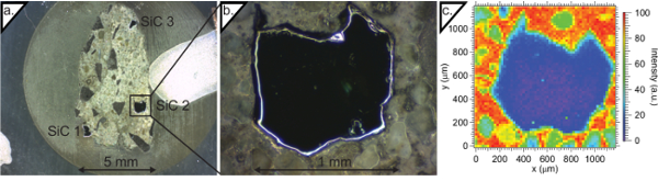 Figure 1: Moissanite sample (a) Moissanite sample embedded in epoxy puck. Three moissanite crystals can be optically identified. (b) Higher magnification microscope image of the region of interest. (c) X-ray fluorescence (XRF) map of the sample. The XRF measures all intensity from 2,000-20,000 eV. Since the Kα1 emission lines ofmore »
Figures / Tables found in this record: