
Two-color monochromatic x-ray imaging with a single short-pulse laser
Abstract
Here, simultaneous monochromatic crystal imaging at 4.5 and 8.0 keV with x-rays produced by a single short-pulse laser is presented. A layered target consisting of thin foils of titanium and copper glued together is irradiated by the 50 TW Leopard short-pulse laser housed at the Nevada Terawatt Facility. Laser-accelerated MeV fast electrons transmitting through the target induce Kα fluorescence from both foils. Two energy-selective curved crystals in the imaging diagnostic form separate monochromatic images on a single imaging detector. The experiment demonstrates simultaneous two-color monochromatic imaging of the foils on a single detector as well as Kα x-ray production at two different photon energies with a single laser beam. Lastly, application of the diagnostic technique to x-ray radiography of a high density plasma is also presented.
- Authors:
-
 [1];
[2];
[1];
[2];
- Univ. of Nevada, Reno, NV (United States). Department of Physics
- Lawrence Livermore National Lab. (LLNL), Livermore, CA (United States)
- Ecole Polytechnique, CNRS (France)
- Publication Date:
- Research Org.:
- Lawrence Livermore National Lab. (LLNL), Livermore, CA (United States)
- Sponsoring Org.:
- USDOE National Nuclear Security Administration (NNSA)
- OSTI Identifier:
- 1458668
- Alternate Identifier(s):
- OSTI ID: 1363707
- Report Number(s):
- LLNL-JRNL-718761
Journal ID: ISSN 0034-6748; 862345; TRN: US1901498
- Grant/Contract Number:
- AC52-07NA27344; FG02-05ER54834; NA0002075
- Resource Type:
- Accepted Manuscript
- Journal Name:
- Review of Scientific Instruments
- Additional Journal Information:
- Journal Volume: 88; Journal Issue: 6; Journal ID: ISSN 0034-6748
- Publisher:
- American Institute of Physics (AIP)
- Country of Publication:
- United States
- Language:
- English
- Subject:
- 70 PLASMA PHYSICS AND FUSION TECHNOLOGY
Citation Formats
                    Sawada, H., Daykin, T., McLean, H. S., Chen, H., Patel, P. K., Ping, Y., and Perez, F. Two-color monochromatic x-ray imaging with a single short-pulse laser.  United States: N. p., 2017.
Web.  doi:10.1063/1.4985729.
Sawada, H., Daykin, T., McLean, H. S., Chen, H., Patel, P. K., Ping, Y., & Perez, F. Two-color monochromatic x-ray imaging with a single short-pulse laser. United States. https://doi.org/10.1063/1.4985729
                    Sawada, H., Daykin, T., McLean, H. S., Chen, H., Patel, P. K., Ping, Y., and Perez, F. Tue .
"Two-color monochromatic x-ray imaging with a single short-pulse laser".  United States.  https://doi.org/10.1063/1.4985729.  https://www.osti.gov/servlets/purl/1458668.
@article{osti_1458668,

  title        = {Two-color monochromatic x-ray imaging with a single short-pulse laser},

  author       = {Sawada, H. and Daykin, T. and McLean, H. S. and Chen, H. and Patel, P. K. and Ping, Y. and Perez, F.},

  abstractNote = {Here, simultaneous monochromatic crystal imaging at 4.5 and 8.0 keV with x-rays produced by a single short-pulse laser is presented. A layered target consisting of thin foils of titanium and copper glued together is irradiated by the 50 TW Leopard short-pulse laser housed at the Nevada Terawatt Facility. Laser-accelerated MeV fast electrons transmitting through the target induce Kα fluorescence from both foils. Two energy-selective curved crystals in the imaging diagnostic form separate monochromatic images on a single imaging detector. The experiment demonstrates simultaneous two-color monochromatic imaging of the foils on a single detector as well as Kα x-ray production at two different photon energies with a single laser beam. Lastly, application of the diagnostic technique to x-ray radiography of a high density plasma is also presented.},

  doi          = {10.1063/1.4985729},

  journal      = {Review of Scientific Instruments},

  number       = 6,

  volume       = 88,

  place        = {United States},

  year         = {Tue Jun 13 00:00:00 EDT 2017},

  month        = {Tue Jun 13 00:00:00 EDT 2017}

}
 Search WorldCat to find libraries that may hold this journal
Web of Science
Figures / Tables:
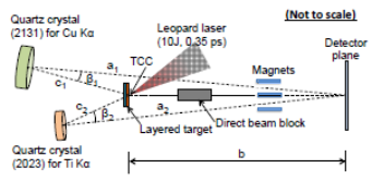 Figure 1: A schematic of two-color monochromatic x-ray imaging with two spherically curved crystals on the Leopard laser experiment (not to scale)
Works referenced in this record:
Development of Compton radiography of inertial confinement fusion implosions
journal, May 2011
- Tommasini, R.; Hatchett, S. P.; Hey, D. S.
- Physics of Plasmas, Vol. 18, Issue 5
The National Ignition Facility and the National Ignition Campaign
journal, April 2010
- Moses, E. I.
- IEEE Transactions on Plasma Science, Vol. 38, Issue 4
Monochromatic x-ray backlighting of wire-array z -pinch plasmas using spherically bent quartz crystals
journal, March 2003
- Sinars, D. B.; Cuneo, M. E.; Bennett, G. R.
- Review of Scientific Instruments, Vol. 74, Issue 3
Systematic search for spherical crystal X-ray microscopes matching 1–25 keV spectral line sources
journal, December 2016
- Schollmeier, Marius S.; Loisel, Guillaume P.
- Review of Scientific Instruments, Vol. 87, Issue 12
Visualizing fast electron energy transport into laser-compressed high-density fast-ignition targets
journal, January 2016
- Jarrott, L. C.; Wei, M. S.; McGuffey, C.
- Nature Physics, Vol. 12, Issue 5
Monochromatic x-ray radiography for areal-density measurement of inertial fusion energy fuel in fast ignition experiment
journal, October 2010
- Fujioka, Shinsuke; Fujiwara, Takashi; Tanabe, Minoru
- Review of Scientific Instruments, Vol. 81, Issue 10
Diagnosis of laser-target implosions by space-resolved continuum absorption x-ray spectroscopy
journal, May 1994
- Marshall, F. J.; Delettrez, J. A.; Epstein, R.
- Physical Review E, Vol. 49, Issue 5
Initial performance results of the OMEGA laser system
journal, January 1997
- Boehly, T. R.; Brown, D. L.; Craxton, R. S.
- Optics Communications, Vol. 133, Issue 1-6
Flash Kα radiography of laser-driven solid sphere compression for fast ignition
journal, June 2016
- Sawada, H.; Lee, S.; Shiroto, T.
- Applied Physics Letters, Vol. 108, Issue 25
High-energy x-ray microscopy techniques for laser-fusion plasma research at the National Ignition Facility
journal, January 1998
- Koch, Jeffrey A.; Landen, Otto L.; Barbee, Troy W.
- Applied Optics, Vol. 37, Issue 10, p. 1784-1795
Time-resolved compression of a capsule with a cone to high density for fast-ignition laser fusion
journal, December 2014
- Theobald, W.; Solodov, A. A.; Stoeckl, C.
- Nature Communications, Vol. 5, Issue 1
X-ray backlighting for the National Ignition Facility (invited)
journal, January 2001
- Landen, O. L.; Farley, D. R.; Glendinning, S. G.
- Review of Scientific Instruments, Vol. 72, Issue 1
Temperature mapping of compressed fusion pellets obtained by monochromatic imaging
journal, January 1995
- Uschmann, I.; Förster, E.; Nishimura, H.
- Review of Scientific Instruments, Vol. 66, Issue 1
10-kJ PW laser for the FIREX-I program
journal, June 2006
- Miyanaga, N.; Azechi, H.; Tanaka, K. A.
- Journal de Physique IV (Proceedings), Vol. 133
High performance compact magnetic spectrometers for energetic ion and electron measurement in ultraintense short pulse laser solid interactions
journal, October 2008
- Chen, Hui; Link, Anthony J.; van Maren, Roger
- Review of Scientific Instruments, Vol. 79, Issue 10
Development of the 50 TW laser for joint experiments with 1 MA z-pinches
journal, August 2010
- Wiewior, P. P.; Ivanov, V. V.; Chalyy, O.
- Journal of Physics: Conference Series, Vol. 244, Issue 3
Temperature sensitivity of Cu Kα imaging efficiency using a spherical Bragg reflecting crystal
journal, February 2007
- Akli, K. U.; Key, M. H.; Chung, H. K.
- Physics of Plasmas, Vol. 14, Issue 2
Time-Resolved Measurements of Hot-Electron Equilibration Dynamics in High-Intensity Laser Interactions with Thin-Foil Solid Targets
journal, February 2012
- Nilson, P. M.; Davies, J. R.; Theobald, W.
- Physical Review Letters, Vol. 108, Issue 8
Soft x-ray backlighting of cryogenic implosions using a narrowband crystal imaging system (invited)
journal, November 2014
- Stoeckl, C.; Bedzyk, M.; Brent, G.
- Review of Scientific Instruments, Vol. 85, Issue 11
Development of backlighting sources for a Compton radiography diagnostic of inertial confinement fusion targets (invited)
journal, October 2008
- Tommasini, R.; MacPhee, A.; Hey, D.
- Review of Scientific Instruments, Vol. 79, Issue 10
Monochromatic focusing of subpicosecond x-ray pulses in the keV range
journal, February 1999
- Missalla, T.; Uschmann, I.; Förster, E.
- Review of Scientific Instruments, Vol. 70, Issue 2
Nd-doped phosphate glass laser systems for laser-fusion research
journal, September 1981
- Yamanaka, C.; Kato, Y.; Izawa, Y.
- IEEE Journal of Quantum Electronics, Vol. 17, Issue 9
4.5- and 8-keV emission and absorption x-ray imaging using spherically bent quartz 203 and 211 crystals (invited)
journal, March 2003
- Koch, J. A.; Aglitskiy, Y.; Brown, C.
- Review of Scientific Instruments, Vol. 74, Issue 3, p. 2130-2135
High-resolution 17–75keV backlighters for high energy density experiments
journal, July 2008
- Park, H. -S.; Maddox, B. R.; Giraldez, E.
- Physics of Plasmas, Vol. 15, Issue 7
Investigating high speed phenomena in laser plasma interactions using dilation x-ray imager (invited)
journal, November 2014
- Nagel, S. R.; Hilsabeck, T. J.; Bell, P. M.
- Review of Scientific Instruments, Vol. 85, Issue 11
X‐ray radiographic measurements of radiation‐driven shock and interface motion in solid density material
journal, July 1993
- Hammel, B. A.; Griswold, D.; Landen, O. L.
- Physics of Fluids B: Plasma Physics, Vol. 5, Issue 7
Performance of bent-crystal x-ray microscopes for high energy density physics research
journal, January 2015
- Schollmeier, Marius S.; Geissel, Matthias; Shores, Jonathon E.
- Applied Optics, Vol. 54, Issue 16
Comprehensive description of the Orion laser facility
journal, April 2015
- Hopps, Nicholas; Oades, Kevin; Andrew, Jim
- Plasma Physics and Controlled Fusion, Vol. 57, Issue 6
Isochoric heating in heterogeneous solid targets with ultrashort laser pulses
journal, December 2007
- Sentoku, Y.; Kemp, A. J.; Presura, R.
- Physics of Plasmas, Vol. 14, Issue 12
Monochromatic x-ray imaging with bent crystals for laser fusion research
journal, January 2001
- Fujita, K.; Nishimura, H.; Niki, I.
- Review of Scientific Instruments, Vol. 72, Issue 1
Absolute measurements of x-ray backlighter sources at energies above 10 keV
journal, May 2011
- Maddox, B. R.; Park, H. S.; Remington, B. A.
- Physics of Plasmas, Vol. 18, Issue 5
Ti Kα radiography of Cu-doped plastic microshell implosions via spherically bent crystal imaging
journal, May 2005
- King, J. A.; Akli, K.; Zhang, B.
- Applied Physics Letters, Vol. 86, Issue 19
Observation of Instability Growth in a Copper $Z$-Pinch Target Using Two-Color Monochromatic X-Ray Backlighting
journal, November 2011
- Sinars, Daniel B.; Peterson, Kyle J.; Slutz, Stephen A.
- IEEE Transactions on Plasma Science, Vol. 39, Issue 11
SPECT3D – A multi-dimensional collisional-radiative code for generating diagnostic signatures based on hydrodynamics and PIC simulation output
journal, May 2007
- MacFarlane, J. J.; Golovkin, I. E.; Wang, P.
- High Energy Density Physics, Vol. 3, Issue 1-2
A dual channel X-ray spectrometer for fast ignition research
journal, July 2010
- Akli, K. U.; Patel, P. K.; Maren, R. Van
- Journal of Instrumentation, Vol. 5, Issue 07
Pulsed-power-driven high energy density physics and inertial confinement fusion research
journal, May 2005
- Matzen, M. Keith; Sweeney, M. A.; Adams, R. G.
- Physics of Plasmas, Vol. 12, Issue 5
X-ray crystal imagers for inertial confinement fusion experiments (invited)
journal, January 1999
- Aglitskiy, Y.; Lehecka, T.; Obenschain, S.
- Review of Scientific Instruments, Vol. 70, Issue 1
Pulse-dilation enhanced gated optical imager with 5 ps resolution (invited)
journal, October 2010
- Hilsabeck, T. J.; Hares, J. D.; Kilkenny, J. D.
- Review of Scientific Instruments, Vol. 81, Issue 10
Progress on converting a NIF quad to eight, petawatt beams for advanced radiography
journal, August 2010
- Crane, J. K.; Tietbohl, G.; Arnold, P.
- Journal of Physics: Conference Series, Vol. 244, Issue 3
Works referencing / citing this record:
Note: A Laue crystal imager for high energy quasi-monochromatic x-ray
journal, September 2018
- Zhang, Zhe; Nishimura, Hiroaki; Yao, Akira
- Review of Scientific Instruments, Vol. 89, Issue 9
Figures / Tables found in this record: