skip to main content
OSTI.GOV title logo U.S. Department of Energy
Office of Scientific and Technical Information

Title: Measuring Three-Dimensional Strain and Structural Defects in a Single InGaAs Nanowire Using Coherent X-ray Multiangle Bragg Projection Ptychography

Journal Article · · Nano Letters
ORCiD logo [1];  [2];  [3];  [4];  [2];  [5]; ORCiD logo [5];  [1];  [6];  [6];  [6];  [6];  [2];  [3]; ORCiD logo [1];  [2]
  1. Northwestern Univ., Evanston, IL (United States). Dept. of Materials Science and Engineering
  2. Argonne National Lab. (ANL), Argonne, IL (United States). Materials Science Division
  3. Aix-Marseille Univ., and CNRS/IN2P3, Marseille (France)
  4. Argonne National Lab. (ANL), Argonne, IL (United States). Center for Nanoscale Materials
  5. Technische Univ. Munich (Germany). Walter Schottky Inst. and Physik Dept.
  6. Brookhaven National Lab. (BNL), Upton, NY (United States). National Synchrotron Light Source II (NSLS-II)

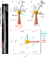
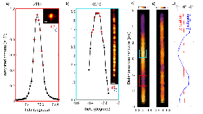
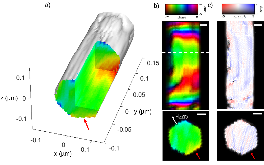
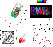
III-As nanowires are candidates for near infrared light emitters and detectors that can be directly integrated onto silicon. However, nanoscale to microscale variations in structure, composition, and strain within a given nanowire, as well as variations between nanowires, pose challenges to correlating microstructure with device performance. In this work, we utilize coherent nano-focused x-rays to characterize stacking defects and strain in a single InGaAs nanowire supported on Si. By reconstructing diffraction patterns from the 2110 Bragg peak, we show that the lattice orientation varies along the length of the wire, while the strain field along the cross-section is largely unaffected, leaving the band structure unperturbed. Diffraction patterns from the 0110 Bragg peak are reproducibly reconstructed to create three-dimensional images of stacking defects and associated lattice strains, revealing sharp planar boundaries between different crystal phases of wurtzite (WZ) structure that contribute to charge carrier scattering. Phase retrieval is made possible by developing multi-angle Bragg projection ptychography (maBPP) to accommodate coherent nanodiffraction patterns measured at arbitrary overlapping positions at multiple angles about a Bragg peak, eliminating the need for scan registration at different angles. The penetrating nature of x-ray radiation, together with the relaxed constraints of maBPP, will enable in operando imaging of nanowire devices.

Research Organization:
Argonne National Lab. (ANL), Argonne, IL (United States); Brookhaven National Lab. (BNL), Upton, NY (United States)
Sponsoring Organization:
USDOE Office of Science (SC), Basic Energy Sciences (BES) (SC-22). Materials Sciences & Engineering Division; European Union (EU); National Science Foundation (NSF); German Research Foundation (DFG); USDOE Office of Science (SC), Basic Energy Sciences (BES)
Grant/Contract Number:
AC02-06CH11357; SC0012704
OSTI ID:
1425485
Alternate ID(s):
OSTI ID: 1426467
Report Number(s):
BNL-203363-2018-JAAM; 138261; TRN: US1802116
Journal Information:
Nano Letters, Vol. 18, Issue 2; ISSN 1530-6984
Publisher:
American Chemical SocietyCopyright Statement
Country of Publication:
United States
Language:
English
Citation Metrics:
Cited by: 43 works
Citation information provided by
Web of Science

References (46)

Growth and properties of InGaAs nanowires on silicon: Growth and properties of InGaAs nanowires on silicon journal December 2013
Monolithic InGaAs Nanowire Array Lasers on Silicon-on-Insulator Operating at Room Temperature journal May 2017
Monolithic III–V on Silicon Plasmonic Nanolaser Structure for Optical Interconnects journal September 2015
Epitaxial III−V Nanowires on Silicon journal October 2004
GaAs–AlGaAs core–shell nanowire lasers on silicon: invited review journal April 2017
A review of III–V nanowire infrared photodetectors and sensors journal February 2017
Structural and optical properties of high quality zinc-blende/wurtzite GaAs nanowire heterostructures journal December 2009
Three-Dimensional Morphology of GaP−GaAs Nanowires Revealed by Transmission Electron Microscopy Tomography journal October 2007
Spontaneous Alloy Composition Ordering in GaAs-AlGaAs Core–Shell Nanowires journal March 2013
Alloy Fluctuations Act as Quantum Dot-like Emitters in GaAs-AlGaAs Core–Shell Nanowires journal July 2015
Correlating the Nanostructure and Electronic Properties of InAs Nanowires journal May 2010
Quantum Transport and Sub-Band Structure of Modulation-Doped GaAs/AlAs Core–Superlattice Nanowires journal July 2017
Luminescence of GaAs nanowires consisting of wurtzite and zinc-blende segments journal January 2012
Crystal Phase Quantum Dots in the Ultrathin Core of GaAs–AlGaAs Core–Shell Nanowires journal October 2015
X-ray Bragg Ptychography on a Single InGaN/GaN Core–Shell Nanowire journal March 2017
Threefold rotational symmetry in hexagonally shaped core–shell (In,Ga)As/GaAs nanowires revealed by coherent X-ray diffraction imaging journal April 2017
Coherent diffraction imaging of a single epitaxial InAs nanowire using a focused x-ray beam journal March 2009
Inversion Domain Boundaries in GaN Wires Revealed by Coherent Bragg Imaging journal September 2015
Determination of the stacking fault density in highly defective single GaAs nanowires by means of coherent diffraction imaging journal June 2016
Characterization of individual stacking faults in a wurtzite GaAs nanowire by nanobeam X-ray diffraction journal August 2017
Widely tunable alloy composition and crystal structure in catalyst-free InGaAs nanowire arrays grown by selective area molecular beam epitaxy journal February 2016
Lattice-Matched InGaAs–InAlAs Core–Shell Nanowires with Improved Luminescence and Photoresponse Properties journal April 2015
High-resolution three-dimensional structural microscopy by single-angle Bragg ptychography journal November 2016
Three-dimensional high-resolution quantitative microscopy of extended crystals journal September 2011
Hard-X-Ray Lensless Imaging of Extended Objects journal January 2007
High-Resolution Scanning X-ray Diffraction Microscopy journal July 2008
Noise models for low counting rate coherent diffraction imaging journal January 2012
3D Imaging of Twin Domain Defects in Gold Nanoparticles journal May 2015
Determination of the cubic to hexagonal fraction in GaN nucleation layers using grazing incidence x-ray scattering journal June 1998
Imaging Local Polarization in Ferroelectric Thin Films by Coherent X-Ray Bragg Projection Ptychography journal April 2013
Nondestructive three-dimensional imaging of crystal strain and rotations in an extended bonded semiconductor heterostructure journal November 2015
On-the-fly scans for X-ray ptychography journal December 2014
Continuous scanning mode for ptychography journal January 2014
Quantitative Nanoscale Imaging of Lattice Distortions in Epitaxial Semiconductor Heterostructures Using Nanofocused X-ray Bragg Projection Ptychography journal September 2012
Role of microstructure on optical properties in high-uniformity In 1 x Ga x As nanowire arrays: Evidence of a wider wurtzite band gap journal May 2013
Optically pumped room-temperature GaAs nanowire lasers journal November 2013
Multimodality hard-x-ray imaging of a chromosome with nanoscale spatial resolution journal February 2016
Analysis of strain and stacking faults in single nanowires using Bragg coherent diffraction imaging journal March 2010
Efficient modeling of Bragg coherent x-ray nanobeam diffraction journal January 2015
Fast Strain Mapping of Nanowire Light-Emitting Diodes Using Nanofocused X-ray Beams journal June 2015
Bragg coherent x-ray diffractive imaging of a single indium phosphide nanowire journal May 2016
Bending and Twisting Lattice Tilt in Strained Core–Shell Nanowires Revealed by Nanofocused X-ray Diffraction journal June 2017
Topological defect dynamics in operando battery nanoparticles journal June 2015
Revealing crystalline domains in a mollusc shell single-crystalline prism journal July 2017
Three Dimensional Variable-Wavelength X-Ray Bragg Coherent Diffraction Imaging journal November 2016
Theoretical analysis of the strain mapping in a single core-shell nanowire by x-ray Bragg ptychography conference September 2015

Cited By (13)

Multimodal X-ray imaging of grain-level properties and performance in a polycrystalline solar cell text January 2019
Unravelling the strain relaxation processes in silicon nanowire arrays by X-ray diffraction journal September 2019
Combining Nanofocused X-Rays with Electrical Measurements at the NanoMAX Beamline journal August 2019
Microscopy Instrumentation and Nanopositioning at NSLS-II: Current Status and Future Directions journal September 2018
Coherent X-ray diffraction imaging meets ptychography to study core-shell-shell nanowires journal January 2018
Ptychographic characterization of a coherent nanofocused X-ray beam journal January 2020
Coherent Bragg imaging of 60 nm Au nanoparticles under electrochemical control at the NanoMAX beamline journal August 2019
X-ray ptychography on low-dimensional hard-condensed matter materials journal March 2019
High performance indium oxide nanoribbon FETs: mitigating devices signal variation from batch fabrication journal January 2019
Three-dimensional optical trapping and orientation of microparticles for coherent X-ray diffraction imaging journal February 2019
Complete structural and strain analysis of single GaAs/(In,Ga)As/GaAs core–shell–shell nanowires by means of in-plane and out-of-plane X-ray nanodiffraction journal September 2018
Multimodal X-ray imaging of grain-level properties and performance in a polycrystalline solar cell journal May 2019
X-ray microscopy journal July 1965

Figures / Tables (4)